Transperineal MRI/US Fusion Biopsy
Transperineal MRI/US Fusion Biopsy
A transperineal prostate biopsy is a procedure used to obtain tissue samples from the prostate gland for diagnostic purposes. Unlike the more common transrectal prostate biopsy, which involves inserting a biopsy needle through the rectum to access the prostate, a transperineal biopsy accesses the prostate through the perineum, which is the area between the scrotum and the anus.
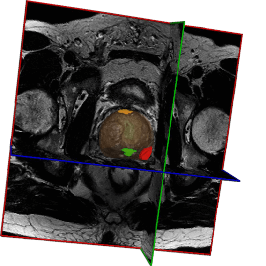
We utilize fusion technology that combines the benefits of MR imaging and real-time ultrasound for our transperineal biopsies. MRI has the advantage of evaluating 100% of the prostate to look for areas suspicious for cancer. Using this information from the MRI, we fuse these findings with the ultrasound machine results. This new fusion information can now target areas that may not be visualized during the standard biopsy.

During a transperineal prostate biopsy, the patient is usually positioned lying on their back with their legs raised and apart. The area between the scrotum and the anus is cleaned and sterilized. Local anesthesia is then administered to numb the area.
A biopsy needle is inserted through the skin of the perineum and into the prostate gland under ultrasound guidance. Multiple tissue samples are then taken from different areas of the prostate using the biopsy needle. The samples are sent to a pathology laboratory for analysis to determine if there are any cancerous cells present in the prostate tissue.
The samples of analysis that are sent to a pathology laboratory:
- Almost zero infection rate.
- No need for antibiotics
- Can be done on the same day (time permitting)
- Cancer detection rate similar
- Able to be performed under local anesthesia
- Do not need to fast overnight prior to the procedure
- Able to walk out of the office
- Enema/suppository is optional
Traditional Transrectal Prostate Biopsy:
- Requires antibiotics which messes up the gut flora
- 5-7% infection rate
- 1-2% sepsis rate
- Requires an enema

